Dr. María Iglesias, Barraquer collaborator, obtains a new award for her LASIK tonometer
12/04/2024
13/02/2018
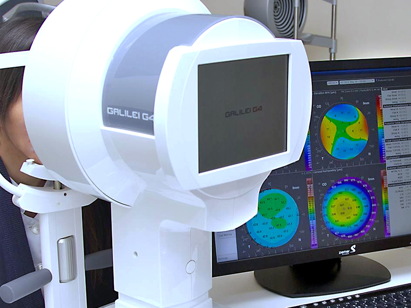
Corneal topography is a non-invasive computerized diagnostic test that creates a 3D map of the surface of the cornea and allows us to know its characteristics.
The cornea is a thin transparent membrane that closes the eyeball, in front of the iris and pupil. It fulfills multiple functions: as a transparent window of the eye it lets the images pass and acts as a powerful lens to focus them on the retina. The retina is a nervous tissue that carries the images to the brain and that allows us to see the objects that surround us.
As in topographic maps, corneal topography is based on level lines in which warm colors represent an elevation and cold colors a depression.
For the patient the test may vary a bit depending on the device but it consists of looking at a fixed point for 3 or 4 seconds, in which you will see a red light or a light sweep that goes from one side to the other.
The information given by the corneal topography is the curvature, thickness, eccentricity ... of the cornea. All this information is relevant for different types of interventions such as corneal refractive surgery, corneal transplantation or cataract extraction, among others. It is also important to know if there are pathologies: keratoconus detection, irregular astigmatism, corneal degeneration, etc. and for the adaptation of contact lenses.