Talking to Dr Rob van der Veen
15/10/2026
19/10/2026
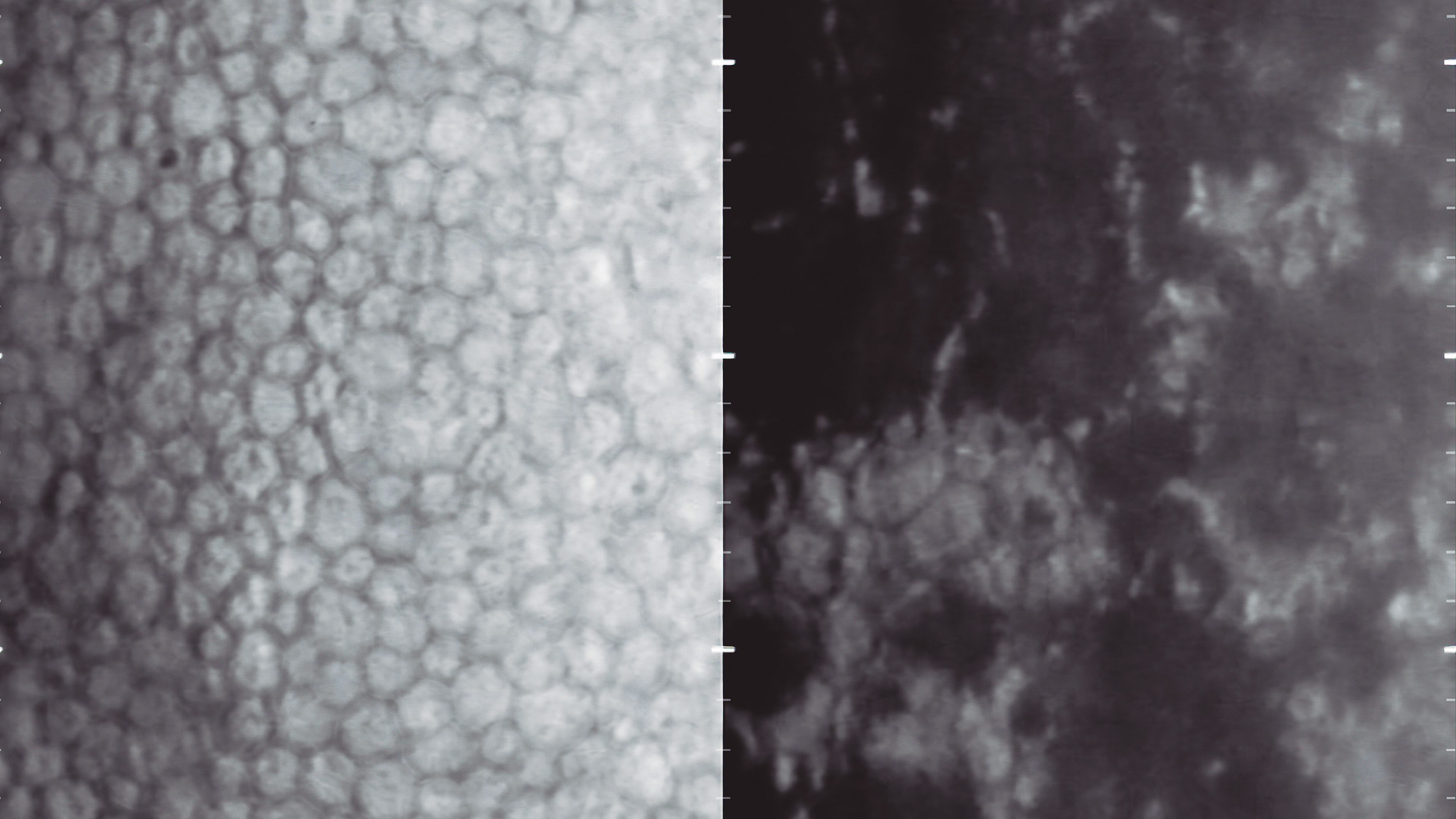
Specular microscopy is an imaging technique that allows detailed examination of the cells of the corneal endothelium, the innermost layer of the cornea. The main function of these cells is to keep the cornea transparent through two main mechanisms:
Unlike other cells in the body, these cells do not regenerate, so their number and shape are key indicators of eye health. When the number of these cells decreases significantly or they do not function properly, the cornea becomes excessively hydrated, leading to corneal oedema, blurred vision and loss of transparency.
It is a quick, painless and non-invasive test. The patient simply rests their chin on the chin rest and focuses on a point of light while the images are captured. Within seconds, the specialist obtains precise information about cell density, cell size and morphology.
One of its main benefits is its ability to anticipate potential complications. Knowing the condition of the endothelium allows the ophthalmologist to tailor treatment to each patient and reduce risks, improving safety and visual outcomes.
In short, specular microscopy is a key tool in modern ophthalmology, as it provides reliable information for the care and preservation of long-term eye health.
Olga Riera, Ocular Biometry Department