Talking to Dr Alberto Lozano
12/06/2026
15/10/2019
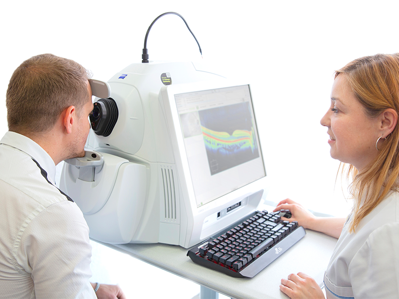
An optical coherence tomography is a non-invasive diagnosis technique which, through obtaining multilple axial sections using light as an energy source, obtains high resolution images that resemble an “in vivo” histological section. This enables pathologies of the retina, choroid and anterior segment to be studied.
A painless and simple test
Most commonly known by its initials in English, an OCT (Optical Coherence Tomography) is a pain-free test. There is no eye contact and there is no need to dilatethe pupil. It’s simple, quick and there are no side effects.
The OCT has become an essential test in daily ophthalmic clinical practice, due to the fact that by using quantitative data, it allows for an objective analysis of structural changes in the morphology and thickness of:
As a result, the OCT improves the efficiency of the diagnosis, follow-up, treatment and the possible surgical approach of the ophthalmologist in retinal pathologies such as age-related macular degeneration (AMD), central serous choroidopathy (CSC), macular holes, epiretinal membranes, occlusion of the central retinal artery occlusion, (CRAO), etc. In glaucoma, it helps us with early detection of the loss of the nerve fibre layer, due to increased ocular pressure, thereby preventing serious and irreversible damage.
The latest innovations in tomographs
The latest generation of tomography equipment like the CIRRUS HD-5000 by ZEISS has retina tracking tools (FastTrack) and an automatic fovea macula centering device (Fovea Finder), helping to gain a more precise analysis of the macular thickness. The new high definition (HD) scans produce images with a higher resolution, thereby improving the quality to help differentiate the retina layers more precisely. The incorporation of the anterior segment module allows us to obtain images of the anterior segment (cornea, iridocorneal angle) and pachymetric thickness measurements of the cornea and endothelium, the layer located in the internal surface of the cornea.
Finally, the Centre’s tomography equipment includes a new feature: the AngioPlex module. It allows us to carry out an Angio-OCT (OCTA). This is a huge leap in the development of OCT technology because it makes obtaining dynamic data on the ocular structures possible by providing information on the vascular flow via red blood cell movement, complementary to the structure information obtained using a conventional OCT.
Raquel Larena, Diagnostic Imaging Department