Ocular inflammation and uveitis as a possible sign of another disease
10/07/2026
22/02/2016
A new technological tool has joined the field of Ophthalmology in the last five years the Femtosecond Laser has increased the safety and precision of certain surgical techniques.
Femtosecond laser
It is a device capable of emitting ultrashort pulses of laser light, with a duration of under one picosecond, in the order of femtoseconds (1fs= 10-15 s). These pulses are emitted at a wavelength of 1053 nm, which allows crossing the transparent tissues of the eye. When they impact, they produce the effect called photodisruption and photoionization, creating a plasma point with a diameter of 1 to 3 microns, an acoustic shock that forms a cavitation micro-bubble separating the tissue. A transparent tissue can be cut when thousands of impacts are positioned one after the other and together at high speed.
Applications
It is currently used in Ophthalmology in corneal and cataract surgery. In the cornea, it is used in transplants and in refractive surgery. With this instrument, we can make penetrating or laminar transplants, with very precise diameter and depth controlled at all times by a display of the cut in real time. This technology was initially used in refractive surgery for the creation of the flap (Fig. 3) in the Laser Assisted In-Situ Keratomileusis (LASIK) technique. Thanks to its application, the complications previously could arise with mechanical cutting tools, such as the microkeratome, have virtually disappeared. The flaps thus obtained have a very precise thickness, which increases the residual corneal thickness obtained after applying the excimer laser, increasing the stability and safety of the procedure. It also allows relaxing arched vertical incisions to correct high astigmatism, make circular tunnels for the implantation of rings in the treatment of diseases such as keratoconus, all with an extremely high precision and in less than half a minute.
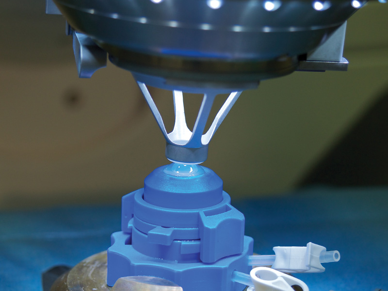
Its use has been developed in cataract surgery in the last five years. With the femtosecond laser, we can make before surgery all necessary cuts to remove the cataract and implant the intraocular lens (Fig. 4). The corneal incisions are made, then a circular cut on the anterior lens capsule and the latter is cut into small fragments that will facilitate their subsequent suction by the surgeon. This laser is used to increase safety and precision in modern cataract surgery.