Why can I have glaucoma if my eye pressure is normal?
30/06/2026
20/04/2015
Optical Coherence Tomography (OCT) is a complementary test with which images are obtained of the tissue to be studied using light as a source of energy. With this technique, we can obtain images very similar to histology sections and analyse the internal characteristics of the part of the eye we wish to study. In order to perform the OCT correctly, light must enter freely. Therefore, intraocular opacities (advanced cataracts, haemorrhages, corneal opacities) impede the correct performance of the test. OCT has basically three areas of clinical ocular application: cornea, glaucoma and retina.
Three areas of application
In corneal applications, among others, it is useful for measuring its thickness and determining the safety margins of refractive surgery.
In the case of retinal applications, these are varied:
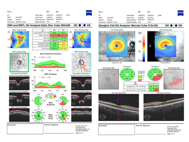
What does OCT allow us to study? (Figure 3 and 4)
In the optic nerve, it allows the measurement of:
In patients with glaucoma, there is a narrowing of the neuroretinal rim as the cup becomes enlarged and the cup-to-disk ratio increases.
In the retinal fibre layer, we can measure the thickness of the nerve fibre layer. In patients with incipient glaucoma, a decrease in said layer can be observed, particularly in the lower and upper quadrants.
In the retinal ganglion cell complex, OCT-Cirrus provides allows accurate measurement thereof in the macula (a decrease in said cells is characteristic of glaucoma).
OCT is a tool for diagnosing incipient glaucoma when there are, as yet, no alterations in the visual field; it also allows us to assess progression and response to the treatment.