How retinal detachment begins and what patients notice
24/07/2026
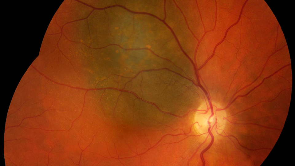

Naevi, also commonly known as moles or freckles, are more or less pigmented lesions that can appear in different parts of the body, especially on the skin and also, although less frequently, at the back of the eye (in a tissue called the posterior uvea or choroid). Although in most cases choroidal naevi do not represent a serious health problem, it is important to know that they require highly specialised medical attention.
Choroidal naevi are pigmented lesions located in the posterior uvea or choroid, a layer of the eye situated beneath the retina.
Although the vast majority of these naevi are benign, some can grow over time. In exceptional cases, there is a risk of transformation into melanoma.
Regular monitoring of a naevus is essential. The ophthalmologist must carry out a thorough examination to rule out clinical signs that could entail a higher potential risk of conversion to a malignant lesion.
Monitoring should include:
The ophthalmologist evaluates:
Based on these findings, the frequency of follow-up will be determined.
According to the international literature, only 0.0005% of choroidal naevi progress to melanoma. However, if several signs of progression appear, it is advisable to carry out:
Cases compatible with melanoma, even small ones, should be treated with very specialised conservative methods:
Choroidal naevi are usually benign lesions, but they require specialised ophthalmic surveillance. At the Barraquer Ophthalmology Centre, we have the technology and experience needed for the diagnosis and follow-up of these lesions, guaranteeing safe and personalised management.
Is it dangerous to have a naevus at the back of the eye?
Not always, but it does require follow-up to detect any change.
Can a naevus at the back of the eye cause pain?
The naevus does not cause pain.
How often should it be checked?
Depending on the associated clinical signs, every 6 to 24 months.
Can naevi turn into melanoma?
Yes, although it is uncommon, and the risk factors for transformation are known: changes in size, pigmentation or the presence of fluid.
What tests do I need?
Examination of the back of the eye (fundus examination), photography, OCT and, sometimes, ultrasound.
Can a benign naevus be operated on?
It is not usually necessary, unless it causes symptoms.
Do naevi appear because of the sun or genetics?
They are not clearly linked to the sun, but there may be a genetic predisposition.
Dr. Javier Elizalde, ophthalmologist at the Barraquer Ophthalmology Centre